
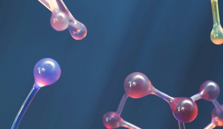
Microcrystal Electron Diffraction (MicroED) is a cutting-edge technique that has revolutionized the field of structural biology. By harnessing the power of electron microscopy, MicroED allows scientists to determine the atomic structure of nanoscale crystals. This article explores the principles behind MicroED, its applications, and its potential impact on various scientific disciplines.
The Basics of Microcrystal Electron Diffraction
MicroED involves the use of a transmission electron microscope (TEM) to analyze tiny crystals, typically in the range of a few hundred nanometers. Unlike traditional X-ray crystallography, which requires larger crystals, MicroED enables the study of smaller crystals that are often challenging to grow. The technique relies on the interaction of electrons with the crystal lattice, producing a diffraction pattern that can be used to determine the arrangement of atoms within the crystal.
Overcoming Challenges in Sample Preparation
One of the key challenges in MicroED is the preparation of suitable samples for analysis. Crystals need to be mounted on a thin support, such as a carbon film, to allow the transmission of electrons. Additionally, the crystals must be oriented in a specific manner to obtain high-quality diffraction patterns. Advances in sample preparation techniques, such as cryo-electron microscopy, have greatly improved the success rate of MicroED experiments.
High-Resolution Structure Determination
MicroED has the potential to provide atomic-level resolution structures of small crystals. By collecting a series of diffraction patterns from different crystal orientations, scientists can solve the phase problem and reconstruct the electron density map. This map can then be used to determine the positions of individual atoms within the crystal. The high resolution of MicroED data allows for the visualization of intricate details, such as hydrogen atoms and solvent molecules, which are often challenging to resolve using other techniques.
Applications in Structural Biology
MicroED has emerged as a powerful tool in the field of structural biology. It has been successfully applied to determine the structures of proteins, peptides, nucleic acids, and small organic molecules. The ability to study small crystals opens up new possibilities for investigating challenging biological targets that were previously inaccessible. MicroED has the potential to contribute to the development of new drugs by providing detailed insights into the interactions between small molecules and their biological targets.
Advancements in Materials Science
Beyond structural biology, MicroED has found applications in materials science. The technique can be used to study the atomic structure of various materials, including catalysts, nanoparticles, and organic-inorganic hybrids. By understanding the precise arrangement of atoms within these materials, scientists can gain insights into their properties and potential applications. MicroED has the potential to accelerate the discovery and development of new materials with tailored properties.
Impact on Drug Discovery
The high-resolution structures obtained through MicroED can significantly impact the field of drug discovery. By visualizing the interactions between drug molecules and their target proteins at the atomic level, scientists can design more effective and specific drugs. MicroED can also aid in the optimization of drug candidates by providing insights into their binding modes and potential modifications. This can potentially lead to the development of safer and more potent drugs.
Conclusion
Microcrystal Electron Diffraction (MicroED) has emerged as a powerful technique for determining the atomic structure of nanoscale crystals. Its applications span across various scientific disciplines, including structural biology and materials science. The ability to study small crystals opens up new possibilities for understanding complex biological processes and designing novel materials. As MicroED continues to evolve, it holds great promise for advancing scientific knowledge and driving innovation.
Citations:
- Nannenga, B. L., Shi, D., Leslie, A. G. W., & Gonen, T. (2014). High-resolution structure determination by continuous-rotation data collection in MicroED. Nature Methods, 11(9), 927–930.
- Rodriguez, J. A., Ivanova, M. I., Sawaya, M. R., Cascio, D., Reyes, F. E., Shi, D., et al. (2015). Structure of the toxic core of α-synuclein from invisible crystals. Nature, 525(7570), 486–490.
- Shi, D., Nannenga, B. L., Iadanza, M. G., & Gonen, T. (2013). Three-dimensional electron crystallography of protein microcrystals. eLife, 2, e01345.
- Yonekura, K., & Gonen, T. (2019). MicroED with the Falcon III direct electron detector. Methods in Enzymology, 632, 155–169.